방선균과 항세균제의 혼용에 의한 과수화상병원균 억제 효과
초록
과수화상병은 사과를 포함한 장미과 식물의 병으로 Erwinia amylovora에 의해 발병하며, 국내에서는 화상병 발병 과수를 땅에 매몰시키는 방제법을 사용하고 있으나 발병이 증가하고 있다. 항세균제와 구리 성분을 대체할 수 있는 생물적 방제법이 연구되고 있으며, 방선균이 유망한 미생물로 알려져 있다. 이 연구는 방선균과 항세균제를 혼용하여 화상병 방제 효과를 증대시키기 위해 수행되었다. 실험 결과, 일부 방선균은 화상병균을 억제하는 효과를 나타내었으며, 항세균제와 혼합 시 효과가 더욱 향상되는 것을 확인했다. 본 연구는 생물적 방제법 개발에 기여할 수 있으며, 화상병의 확산을 통제하는 데 도움이 될 수 있을 것으로 사료된다.
Abstract
This study examined the efficacy of an integrated approach for management of fire blight disease caused by the Gram-negative bacterium Erwinia amylovora. Fire blight, characterized by severe necrosis, poses a significant threat to plants in the Rosaceae family, including apples and pears. Despite efforts that include the use of chemical control, antibiotics, and copper-based pesticides, the pathogen’s high contagion rate poses a barrier to containment. In addition, along with the emergence of antibiotic resistance there are also environmental concerns associated with use of copper-based pesticides. To address these challenges, we examined the potential for combining antagonistic Actinomycetes with bactericides to enhance the inhibitory effects while minimizing the potential for emerging resistance. Of the seven Actinomycetes strains tested, five strains exhibited tolerance to bactericides. Furthermore, the results of combined treatment on plant tissues showed enhanced inhibition compared with individual treatments. These findings provide evidence of the utility of antagonistic bacteria as a complement to chemical control, offering a promising strategy for effectively suppressing E. amylovora and mitigating the development of resistance, thus supporting a comprehensive approach to management of fire blight.
Keywords:
Actinomycetes, Biological control, Combination applications, Erwinia amylovora키워드:
방선균, 생물적 방제, 혼합처리, 과수 화상병서 론
사과는 장미과 사과나무속(Malus)에 속하는 식물로, 우리나라에서 생산량이 높은 과일 중 하나이다(Francini et al., 2022). 사과는 식이 섬유와 비타민C, 미네랄 등 생리 활성물질을 다량 함유하고 있어 영양학적으로 높은 가치를 가지고 있다고 평가된다(Francini et al., 2022). 화상병(Fire blight)은 사과를 포함한 배, 모과 등이 속하는 장미과 식물에 발병하는 주요 병해 중 하나로, 그람 음성 세균인 Erwinia amylovora에 의해 발병하는 병이다. 과수화상병(E. amylovora)는 잎, 줄기, 꽃 등을 통해 감염되어 식물이 불에 그을린 듯 검은 변색을 시작으로 잎맥을 따라 퍼져 검거나 붉게 변하며, 식물체 감염부위에서 외부로 분비되는 세균 누출액(ooze)이 빗물이나 곤충을 통해서 주변 과수에 전파된다(Slack et al., 2017). 국내에서는 2015년에 안성, 천안, 제천의 43개 농가에서 처음 보고된 이래로 현재까지 발생 지역이 꾸준히 확산되고 있다(Ham et al., 2020). 전파력이 매우 빠르고 강하며 경제적 손실이 크기 때문에, 한국에서는 화상병 종합 관리 대책 지침에 따라 화상병 발생과원은 기주식물의 매몰 및 3년간 재배 금지를 실시하고 있다(Kim et al., 2019a). 또한 발병 과원으로부터 반경 5 km 이내의 구역을 정기적으로 예찰 및 연 2회의 화학적 방제를 실시하였으나, 이러한 노력에도 불구하고 2019년 이후로 화상병의 발병이 급격히 증가하였다(Ham et al., 2019).
화상병은 전염력이 매우 높아 과거부터 방제에 어려움을 겪어 왔으며, 항세균제 계열 농약의 장기간 사용으로 인해 화상병원균의 약제 저항성의 발생이 보고되었다(Tancos et al., 2016). 화상병 방제에 50년이 넘는 기간 동안 미국에서 사용되어 왔던 항세균제 streptomycin은 오남용으로 인해 1970년도부터 미국에서는 화상병원균의 항세균제 저항성이 확산된 것으로 보고되었으며, 이로 인하여 화상병 방제에 어려움을 겪었다(Tancos et al., 2016). 또한 최근 환경 오염문제가 대두되는 가운데, 살균제에 주로 사용되던 구리 성분이 포함된 농약의 잔류성 및 독성에 대한 우려 또한 커지고 있다(Aćimović et al., 2015; Dagher et al., 2020).
이러한 한계점에도 불구하고 항세균제 및 구리 성분의 농약을 대체할 만큼 높은 화상병 방제 효과를 보이는 관리방법이 없었기 때문에, 여러 종류의 살균제를 혼용하여 최소한의 기간 내에서 화상병을 방제하도록 규정하고 있다. 화학적 방제를 대체 또는 보완하고자 제시된 접근법으로 병원균에 대한 항세균력 및 병원성을 무력화시키는 기능을 가지는 미생물을 활용하는 생물적 방제 방법이다. 화학적 방제보다 효과가 약하고 느리게 나타나지만, 친환경적이고 저항성 병원균의 출현에 있어서 안전하다는 장점을 가지고 있다. 현재까지 과수 화상병의 생물적 방제제로 알려진 균으로 Pseudomonas fluorescens EPS62e (Bonaterra et al., 2007; Cabrefiga et al., 2011), Pantoea agglomerans E325 (Pusey and Wend, 2012), 및 Lactobacillus plantarum PM411(Daranas et al., 2018) 등이 연구되어 왔다. 방선균은 이러한 생물적 방제법에 활용되는 대표적인 미생물로, 방선균문(Actinobacteria)에 속하는 세균을 총칭한다. 방선균은 다른 종류의 세균류와 달리 균사로 생장하며, 외생 포자를 생성하는 특징이 있는 그람 양성균이다(Kim et al., 2019b). 방선균은 주로 식물의 근권에서 많이 서식하며, 병원균의 생장을 억제시키는 chitinases, glucanases, amylases, cellulases, lipases와 protease 등의 효소나 항생물질을 포함한 다양한 2차 대사산물을 생성하고, 여러 효소 활성에 의해 식물의 성장을 자극시키는 효과 또한 나타나는 것으로 알려져 있다(Jog et al., 2016; Ebrahimi-Zarandi et al., 2022). 본 연구에서는 화학 항세균제를 사용하되 항세균성 방선균을 함께 처리하여, 과수 화상병 방제의 효과 극대화에 대한 기초 정보를 마련하고자 수행되었다. 연구의 목적은 약효가 느리게 나타나는 미생물 농약의 단점과, 저항성 미생물의 출현 가능성이 있는 살균제의 단점을 함께 혼용함으로써 최소화하며 화상병의 확산을 통제하고자 함이다.
재료 및 방법
방선균 및 화상병원균 배양 조건
본 연구에서 평가된 총 7 균주(HJ2F8, J6, KPB2, S8, SN1G12, SP6C4, W1SF4)의 방선균은 전장유전체 분석 등 다양한 선행연구에서 균주의 특징이 확인된 우수한 균주를 화상병 방제 약제와의 혼용성을 평가하였다(Kim et al. 2019b). 방선균은 ISP4 agar media (International Streptomyces Project Medium 4; soluble starch, 10 g; MgSO4·7H2O, 1 g; NaCl, 1 g; (NH4)2SO4, 2 g; CaCO3, 2 g; trace salts solution (FeSO4·7H2O, 0.1 g; MnCl2·4H2O, 0.1 g; ZnSO4 × 7H2O, 0.1 g per 100 mL), 1 mL; agar, 20 g per L, pH 7.2)에서 7일간 배양되었다. 화상병균(Erwinia amylovora TS3128)은 MGY media (Mannitol Glutamate Yeast extract; D-mannitol, 10 g; L-glutamic acid, 2 g; KH2PO4, 0.5 g; NaCl, 0.2 g; MgSO4·7H2O, 0.2 g; yeast extract, 1 g; agar, 15 g per L, pH 7.0)를 이용하여 배양하였다.
방선균의 과수 화상병원균 성장 억제 효과
방선균 균주들의 화상병균에 대한 항세균력을 조사하기 위해서 대치배양을 실시하였다. 멸균된 paper disc (8 mm)를 1/5 PDK (potato dextrose[Difco], 10 g; peptone, 10 g; agar, 20 g per L)에 치상 후, OD600에서 0.7로 희석한 방선균을 20 μL 분주하여 28oC에서 3일간 정치 배양하였다. MGY media에서 2일 배양한 E. amylovora를 원심분리기(8,000 rpm, 10 min)를 이용하여 세균을 회수 후 멸균수로 2번 정제하였다. 이후 OD600에서 0.4로 희석하여 0.2% Carageenan과 1:1로 희석하고, 방선균이 배양된 배지 위에 1 mL 분주 도말하여 3일간 배양하였다. 이후 방선균 주변에 병원균의 생장 억제 여부 및 항세균력 활성을 평가하였다.
방선균의 항세균제 감수성 검정
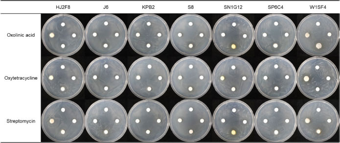
과수 화상병 방제에 주로 사용되는 화학 농약으로 살균제 성분 3종 Streptomycin (a.i. 20%), Oxytetracycline (a.i. 34%), Oxolinic acid (a.i. 20%)에 대해 방선균 균주의 약해 여부를 평가하였다. 방선균 현탁액을 OD600 0.3으로 조정후 ISP3 media (white oats, 20 g; ferric sulphate heptahydrate, 0.001 g; manganese chloride tetrahydrate, 0.001 g; zinc sulphate heptahydrate, 0.001 g; agar, 18 g per L, pH 7.3 ± 0.2)에 100 μL 분주 후 도말하였다. 이후 4방향으로 paper disc를 올려, 화상병에 사용되는 살균제의 기준 사용약량의 0, 1/100, 1/10, 1 농도로 희석한 항세균제를 20 uL 분주하였다. 이 때 항세균제 농도는 시계방향으로 낮은 농도에서 높은 농도로 분주하여 약제 감수성을 확인하였다.
방선균과 살균제 혼용 시 항세균력 상승 효과 검증(In vivo)
방선균 균주들과 항세균제를 함께 사용했을 때 화상병원균에 대한 억제력 및 미생물의 생장이 향상되는지를 확인하기 위해 1/5 PDK에 O.D.600 0.2의 화상병균을 100 μL 도말 후, 4방향으로 8 mm paper disc를 치상하였다. 이후 4방향의 paper disc에 각각 항세균제 20 μL, 방선균 20 μL, 방선균 20 μL와 항세균제 20 μL 혼합, 멸균수 20 μL을 분주하였다. 이후 28oC 항온기에서 3일 간 정치배양 후, paper disk 주변 E. amylovora의 생장 억제를 평가하였다.
사과 유과를 이용한 방선균과 살균제 혼용 상승 효과 검증(In planta)
방선균과 항세균제의 상승 효과가 식물체 내에서 작용하는지를 확인하고자 사과의 유과에서 미생물과 살균제의 항세균력 시너지 효과를 실험을 통해 확인하였다. 사과 유과는 23년 5월 11일 거창 소재의 경상남도 농업기술원 사과이용 연구소에서 직경 1~2 cm의 후지 품종을 채취하였다. 1% NaOCl에서 30초 간 표면을 소독하여 멸균수로 2번 세척 후, 반으로 절단하여 볼록한 면의 중앙에 지름 7 m m 코르크로 표면에 구멍을 뚫었다. 처리구는 항세균제에 약해가 없는 방선균 5종(HJ2F8, J6, KPB2, SP6C4, S8)과 항세균제 3종(Streptomycin, Oxytetracycline, Oxolinic acid)을 단용, 혼용으로 나누어 30 반복으로 진행하였다. 방선균은 모두 OD600 0.3으로 농도를 고정하여 50 μL 분주하였고, 항세균제는 사용약량 농도로 50 μL 분주하였다. 약제 처리 24시간 후에 화상병균(OD600 0.2)을 20 μL를 분주하여 28oC에서 7일 후 방제효과를 확인하였다.
발병도(Disease index, DI)를 결정하는 기준은 접종 부위 주변의 수침상 정도, 접종 부위 주변의 갈변화 정도로 나누어 조사하였다(Fig. 1). 발병도는 병징 정도를 0부터 5로 나누어 0: 어떠한 병징도 보이지 않음, 1: 수침상 1~5%, 갈변화 1~3%, 2: 수침상 6~15%, 갈변화 4~10%, 3: 수침상 16~50%, 갈변화 11~30%, 4: 수침상 51~70%, 갈변화 31~60%, 5: 수침상 71~100%, 갈변화 61~100%로 설정하여 조사하였다(Lee et al., 2018). 이후 병징 정도를 기준으로 발병도를 평가하였다(Safi et al., 2020).

Disease index of fire blight in young apple fruit. 0: no symptoms, 1: water soak 1-5%, browning 1-3%, 2: water soak 6-15%, browning 4-10%, 3: water soak 16-50%, brown Change 11-30%, 4: water soaking 51-70%, browning 31-60%, 5: water soaking 71-100%, browning 61-100%. Afterward, the degree of disease was evaluated based on the degree of symptoms (Lee et al., 2018).
조사 후 방제 효과의 통계적 유의성 확인은 Kruskal-Wallis Rank Sum Test를 진행하여 검증하였다. 이후 Conover-Iman test 사후분석을 진행하였으며, bonferroni로 p-value를 보정하였다. 모든 그래프와 유의값들은 R program(version 4.0.3)의 ggplot2 package를 사용하여 시각화 하였다. 화상병원균을 사용하는 모든 실험은 국립농업과학원에서 실시하였다.
결과 및 고찰
방선균의 화상병원균 억제력 및 살균제 감수성 검정
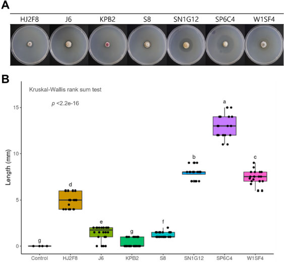
과수 화상병의 화학 항세균제와 상승효과를 가지는 방선균을 선발하기 위하여, 본 실험에 사용한 방선균은 7가지 HJ2F8, J6, KPB2, S8, SN1G12, SP6C4, W1SF4 균주가 사용되었다(Fig. 2A). 해당 균주들을 이용하여 E. amylovora에 대한 항세균능을 검정한 결과, SP6C4의 병원균 생장 억제의 반지름 길이가 평균 12.95 mm로 가장 큰 것을 확인하였다. SP6C4의 병원균 생장 억제 효과가 가장 우수하였으나, 다른 균주들에 비해 생장 저지 구역이 뚜렷하지 못하고 생장억제 부위 내부에 화상병균이 낮은 밀도로 자란 것이 확인되었다. SN1G12 (평균 7.9 mm), W1SF4 (평균 7.41 mm), HJ2F8 (평균 4.975 mm)가 그 다음으로 화상병균에 대하여 높은 항세균성을 보였으며, J6 (평균 1.325 mm), S8 (평균 1.3 mm), KPB2 (0.2 mm)는 반지름 평균이 1.5 mm 이하의 상대적으로 저조한 생장 억제 효과를 나타내었다(Fig. 2B).

Disease severity in inhibitory activity assays against E. amylovora was examined in young apple fruits

(A) Antimicrobial activity of actinobacteria against Erwinia amylovora by well disc diffusion method. Actinobacteria concentration was the same as O.D.600 0.7, O.D.600 0.4 Erwinia amylovora was mixed with 0.2% carrageenan in a 1:1 ratio to measure the clean zone length was measured after 7 days of overlay. (B) Data were analyzed by Kruskal-Wallis rank sum test followed by the Conover post-hoc test for statistical analysis between actinomycetes. Different letters show statistically significant differences (p<0.05).
실험에 사용된 후보 방선균의 생장이 화상병균용 항세균제에 대한 감수성을 평가하였다. 총 7종 중, 5종의 방선균(HJ2F8, J6, KPB2, S8, SP6C4)은 모두 항세균제에 의해 생장이 억제되지 않았으므로, 혼용 처리가 가능할 것이라 판단되었다. 2종의 방선균은 항세균제에 감수성으로 평가되었으며, SN1G12는 streptomycin, W1SF4는 oxytetracycline 성분이 희석농도 1/10 이상에서 각각 억제됨을 확인하였다(Fig. 3). 이후 해당 2종은 살균제와의 혼용 처리가 적합하지 않다고 판단되어 유과에서의 살균제 혼용 실험에서 제외하고 수행하였다.
방선균과 살균제 혼용에 따른 화상병균의 억제 효과
항세균제에 대한 감수성 평가 결과에서 항세균제에 의해 억제되지 않는 5종의 방선균이 항세균제와 혼용되었을 때, 효과적으로 화상병균의 억제력을 향상시킬 수 있는지 확인하기 위해 plate와 사과 유과, 두 가지 환경에서 혼용효과 검증을 진행하였다. 먼저 plate 상에서의 상승 효과를 확인한 결과, 방선균 단독으로 사용했을 때, 방선균 처리농도 OD600 0.7 대비(Fig. 2), OD600 0.3의 농도에서는 E. amylovora에 대한 항세균력을 거의 보이지 않았다(Fig. 4). 이는 방선균의 항세균력을 유도하는 2차 대사산물이 방선균의 성장정체기 또는 포자생성 시기에 생산되기 때문이라 판단된다(Ruiz et al., 2010). 따라서 방선균이 높은 활성을 나타내기 위해서는 포자생성시기에 처리 또는 일정 이상의 밀도를 유지하며 군집화 되는 것이 매우 중요하다고 사료된다.

Synergetic inhibitory effect of antibiotics observed on plates in the presence of microbial co-culture. 20 μL of diluted water on the top, 20 μL of actinomycetes with a density of O.D.6000.3 on the left, 20 μL of bactericide adjusted to the standard use density used in E. amylovora on the right, and 20 μL of actinomycetes and 20 μL of bactericide on the bottom on a paper disk.
살균제 단독 분주 시 화상병원균의 생장억제 평균 길이는 oxolinic acid는 2.25 mm, oxytetracycline는 1.28 mm, streptomycin은 0.27 mm를 나타냈다(Fig. 5). Oxolinic acid와 방선균을 혼합한 경우, 병원균 억제 평균 길이가 SN1G12, 6.25 mm; SP6C4, 6.25 mm; S8, 5.75 mm; KPB2, 5.625 mm; HJ2F8, 5.38 mm; W1SF4, 5 mm 그리고 J6, 4.87 mm 순으로 높은 억제력을 보였다. 대부분의 방선균이 oxolinic acid와 함께 처리 시 약해 현상이 관찰되지 않고, 화상병균에 대한 항세균력이 유의미하게 향상되는 것을 확인하였다(Fig. 5A). 또한 oxytetracycline과 방선균을 혼용한 경우에도 SP6C4, 4.125 mm; S8, 4 mm; HJ2F8, 3.875 mm; J6, 3.625 mm; SN1G12, 3.375 mm; KPB2, 2.5mm로, Oxytetracycline 단독 사용 시 평균 길이가 1.28 mm 인 것에 비하여 평균적으로 2배 이상의 화상병원균에 대한 억제 능력이 상승하는 결과가 관찰되었다(Fig. 5B). SN1G12의 경우, oxytetracycline 단용 시와 혼용 시 간의 억제력이 통계적으로 유의한 차이가 없다는 것을 확인하였으나, 방선균 단용, oxytetracycline 단용, 방선균과 oxytetracycline 혼용 순으로 화상병원균에 대한 억제 효과가 증가하는 것을 알 수 있었다. Streptomycin의 경우 과수화상병을 방제할 때 이용되는 기준 사용약량과 동일한 농도를 사용했음에도 불구하고 화상병원균의 생장 억제효과가 나타나지 않았으나, HJ2F8, J6, S8을 제외한 모든 균주와 혼용 시 병원균 억제 길이가 약 1 mm에서 2 .5 m m까지 증가하는 것을 확인하였다(Fig. 5C). 방선균 단독으로 처리하였을 경우에는 대부분의 방선균들이 OD600 0.3의 밀도에서 화상병원균에 대한 항세균능을 보이지 않는 반면, 항세균제와의 혼용 시 항세균제 단독 사용의 경우보다 E. amylovora의 억제력이 향상되는 효과를 보였다. 이는 곧 미생물과 살균제와의 혼용 시 기대할 수 있는 효과 중 하나로, 살균제의 항생 성분에 의해 억제되지 않는 동시에 억제력을 증진시킬 수 있을 것이라 기대된다.

Synergetic inhibitory effect on E. amylovora by mixed use of bactericides and actinomycetes on a plate. The bactericides used were (A) Oxolinic acid, (B) Oxytetracycline, and (C) Streptomycin, respectively. 20 μL of sterilized water, 20 μL of Actinomycetes with an O.D.600 of 0.3, 20 μL of bactericides, and 20 μL of Actinomycetes and 20 μL of bactericides were added to each of the four paper disks, and the concentration of bactericides was used based on the amount of pesticide used for fire blight. Data were analyzed by Kruskal–Wallis rank sum test followed by the Conover post-hoc test for statistical analysis between treatments. P-value ns: P > 0.05, *: P ≤ 0.05, **: P ≤ 0.01, ***: P ≤ 0.001.
방선균과 항세균제의 혼용처리에 의한 상승효과가 식물체에서도 효과적인지 검증하기 위하여, 해당 실험을 사과의 유과를 이용하여 평가를 진행하였다. 사과 유과에서 E. amylovora의 접종에 의해 화상병의 발병도가 84.7%인 반면, 살균제 단용 처리 시 9~18%까지 발병도가 감소하는 것을 확인하였다(Fig. 6). 사과 유과 접종실험 결과, 배지상에서의 항세균력 실험 결과와 유사하게 streptomycin의 억제력이 가장 낮은 것을 확인하였다. 현재 한국에서 발병한 화상병원균은 전장 유전체 분석 결과 북미의 E. amylovora와 99.5~99.98% 이상의 유사성을 보였기 때문에, 북미의 streptomycin 내성을 가지는 균주가 한국에 도입됐을 가능성을 시사하는 결과로 추론된다(Song et al., 2021). 방선균 단용 효과는 KPB2, 5.3%; J6, 6.7%; HJ2F8, 7.3%; S8, 16.7%; SP6C4, 24.7%로 항세균제 단용 효과보다는 낮은 억제력을 보였다. SP6C4 방선균 단독 접종 시 배지상에서는 가장 우수한 화상병 억제 효과를 보였음에도 불구하고, 유과에서 상대적으로 낮은 억제 효과를 보였다(Fig. 6). 이는 배지상의 억제구역 내부의 E. amylovora가 완전히 저해되지 않은 결과와 비슷한 양상을 보여준다.

Differences in inhibitory activity against E. amylovora between mono- and mixed-use fungicides and actinomycetes in young apple fruits. After cutting the young apple fruits in half, a hole was pierced using a 0.7 mm cork in a round area. Then, 50 μL of Actinomycetes with an O.D.600 of 0.3 and 50 μL of bactericide were dispensed. One day later, 20 μL of Erwinia amylovora at O.D.600 of 0.2 was dispensed, and antibacterial activity was confirmed. P-value ns: P > 0.05, *: P ≤ 0.05, **: P ≤ 0.01, ***: P ≤ 0.001.
방선균과 항세균제의 단독 처리 보다 혼용 시 화상병의 발병률이 감소하는 것을 확인하였다. Oxolinic acid의 경우 단용 시 발병도가 9.3%인데 비하여 혼용 시 J6, 0%; KPB2, 1.3%; S8, 2.6%; SP6C4, 4.7%; HJ2F8, 5.3% 순으로 발병도가 감소하였다. 하지만, HJ2F8, SP6C4, S8는 oxolinic acid 단용과 방선균, oxolinic acid 혼용 간 억제 효과가 통계적으로 차이가 나지 않았다. Oxytetracycline의 단용 효과는 발병도 11.3%인 것에 비하여, 방선균과 Oxytetracycline 혼용 시 KPB2, 1.3%; SP6C4, 1.3%; HJ2F8, 2.7%; J6, 5.3%; S8 8.6% 순으로 발병 억제효과가 증가했으며, Streptomycin의 단용 효과는 18%인 반면, SP6C4, 0.7%; HJ2F8, 1.3%; S8, 1.3%; J6, 2%; KPB2, 6% 순으로 억제 효과가 증가하였다. 결과적으로 화상병 대응 항세균제중 streptomycin이 방선균과 혼용했을 때 발병도가 유의미하게 감소하는 가장 큰 상승작용 효과를 보였으며, 살균제 단용 시 Oxolinic acid와 Oxytetracycline 보다 streptomycin의 방제 효과가 가장 낮았던 것에 비해 KPB2를 제외한 방선균에서 streptomycin을 혼용했을 때, 화상병의 발병이 가장 우수하게 억제되는 것을 확인하였다. 이러한 결과를 통해서 유용미생물의 생장에 영향을 주지 않는 동시에 혼용 시 과수 화상병의 억제 효과가 증대되는 특정 항세균제 및 방선균 조합이 존재한다는 것을 확인할 수 있다. 특히 streptomycin의 경우, 한국에서 화상병의 방제에 많이 사용되는 대표적인 공급약제로, 저항성 균주가 출현했을 때 나타나는 경제적 손실은 아주 클 것이라고 판단된다. 따라서 streptomycin과 선발된 방선균의 혼용을 통해서 더 확실한 병원균 및 병 확산을 억제하고, 저항성 병원균의 출현을 예방할 수 있을 것이라 예상된다.
Acknowledgments
본 연구는 농촌진흥청 연구과제(과제번호: RS-2020-RD009282)의 지원에 의해 수행되었습니다.
이해상충관계
저자는 이해상충관계가 없음을 선언합니다.
References
- Aćimović SG, Zeng Q, McGhee GC, Sundin GW, Wise JC, et al., 2015. Control of fire blight (Erwinia amylovora) on apple trees with trunk-injected plant resistance inducers and antibiotics and assessment of induction of pathogenesisrelated protein genes. Front. Plant Sci. 6:16 DOI 10.3389/ fpls.2015.00016
- Anusha BG, Naik MK, Gopalakrishnan S, Amaresh YS, Gururaj S, 2019. A review on the biological control of plant diseases using various microorganisms. J. Pharmacogn. Phytochem. 8(2):506-510.
-
Bonaterra A, Cabrefiga J, Camps J, Montesinos E, 2007. Increasing survival and efficacy of a bacterial biocontrol agent of fire blight of rosaceous plants by means of osmoadaptation. FEMS Microbiol. Ecol. 61(1):185-195.
[https://doi.org/10.1111/j.1574-6941.2007.00313.x]

-
Cabrefiga J, Francés J, Montesinos E, Bonaterra A, 2011. Improvement of fitness and efficacy of fire blight biocontrol agent via nutritional enhancement combined with osmoadaptation. Appl. Environ. Microbiol. 77(10).
[https://doi.org/10.1128/AEM.02760-10]

- Dagher F, Olishevska S, Philion V, Zheng J, Deziel E, 2020. Development of a novel biological control agent targeting the phytopathogen Erwinia amylovora. Heliyon 6: e05222. DOI 10.1016/j.heliyon.2020.e05222
- Daranas N, Badosa E, Francés J, Montesinos E, Bonaterra A, 2018. Enhancing water stress tolerance improves fitness in biological control strains of Lactobacillus plantarum in plant environments. PLoS One 13(1):e0190931. DOI 10. 1371/journal.pone.0190931.
- Ebrahimi-Zarandi M, Saberi Riseh R, Tarkka MT, 2022. Actinobacteria as effective biocontrol agents against plant pathogens, an overview on their role in eliciting plant defense. Microorganisms 10(9):1739. DOI 10.3390/microor ganisms10091739
- Francini A, Fidalgo-Illesca C, Raffaelli A, Sebastiani L, 2022. Phenolics and mineral elements composition in underutilized apple varieties. Horticulturae 8(1):40. DOI 10.3390/horti culturae8010040
-
Ham HH, Lee KJ, Hong SJ, Kong HG, Lee MH, et al., 2019. Outbreak of fire blight of apple and pear and its characteristics in Korea in 2019. Res. Plant Dis. 26(4): 239-249. (In Korean)
[https://doi.org/10.5423/RPD.2020.26.4.239]

-
Ham HH, Lee YK, Kong HG, Hong SJ, Lee KJ, et al., 2020. Outbreak of fire blight of apple and Asian pear in 2015- 2019 in Korea. Res. Plant Dis. 26(4): 222-228. (In Korean)
[https://doi.org/10.5423/RPD.2020.26.4.222]

-
Jog R, Nareshkumar G, Rajkumar S, 2016. Enhancing soil health and plant growth promotion by actinomycetes. pp. 33-45. In: Subramaniam G, Arumugam S, Rajendran V (Eds.). Plant Growth Promoting Actinobacteria. Springer, Singapore.
[https://doi.org/10.1007/978-981-10-0707-1_3]

-
Kim YE, Kim JY, Noh HJ, Lee DH, Kim SS, et al., 2019a. Investigating survival of Erwinia amylovora from fire blight-diseased apple and pear trees buried in soil as control measure. Korean J. Environ. Agric. 38(4):269-272. (In Korean)
[https://doi.org/10.5338/KJEA.2019.38.4.36]

- Kim DR, Cho GJ, Jeon CW, Weller DM, Thomashow LS, et al., 2019b. A mutualistic interaction between Streptomyces bacteria, strawberry plants and pollinating bees. Nat. Commun. 10(4802). DOI 10.1038/s41467-019-12785-3
-
Lee MS, Lee I, Kim SK, Oh CS, Park DH, 2018. In vitro screening of antibacterial agents for suppression of fire blight disease in Korea. Res. Plant. Dis. 24(1):41-51. (In Korean)
[https://doi.org/10.5423/RPD.2018.24.1.41]

-
Pusey PL, Wend C, 2012. Potential of osmoadaptation for improving Pantoea agglomerans E325 as biocontrol agent for fire blight of apple and pear. Biol. Control 62(1):29-37.
[https://doi.org/10.1016/j.biocontrol.2012.03.002]

-
Ruiz B, Chavez A, Forero A, Garcia-Huante Y, Romero A, et al., 2010. Production of microbial secondary metabolites: Regulation by the carbon source. Crit. Rev. Microbiol. 36(2): 146-167.
[https://doi.org/10.3109/10408410903489576]

-
Safi H, Hussain S, Shahid M, Nazir M, 2020. Incidence and severity of early blight of tomato in Peshawar, Mardan and Malakand divisions and variability amongst the isolates of Alternaria solani jones and mart. Int. J. Agric. Environ. Biotechnol. 13(2):175-183.
[https://doi.org/10.30954/0974-1712.02.2020.9]

-
Slack SM, Zeng Q, Outwater CA, Sundin GW, 2017. Microbiological examination of Erwinia amylovora exopolysaccharide ooze. Phytopatholgy 107(4):403-411.
[https://doi.org/10.1094/PHYTO-09-16-0352-R]

-
Song JY, Yun YH, Kim GD, Kim SH, Lee SJ, et al., 2021. Genome analysis of strains responsible for a fire blight outbreak in Korea. Plant Dis. 105(4):1143-1152.
[https://doi.org/10.1094/PDIS-06-20-1329-RE]

-
Tancos KA, Villani S, Kuehne S, Borejsza-Wysocka E, Breth D, et al., 2016. Prevalence of streptomycin-resistant Erwinia amylovora in New York apple orchards. Plant Dis. 100(4): 802-809.
[https://doi.org/10.1094/PDIS-09-15-0960-RE]

Yejin Lee, Division of Applied Life Science (BK21Plus), Gyeongsang National University, Master student, https://orcid.org/0000-0002-2004-5092
Youn-Sig Kwak, Research Institute of Life Science, Gyeongsang National University, Professor, http://orcid.org/0000-0003-2139-1808

